SBPMD Histology Laboratory Manual
Connective Tissue: Reticular
Reticular tissue, a type of loose connective tissue in which reticular fibers are the most prominent fibrous component, forms the supporting framework of the lymphoid organs (lymph nodes, spleen, tonsils), bone marrow and liver. Reticular fibers (Type III collagen) are too thin to stain in ordinary histological preparations, but they are readily demonstrated by techniques involving the reduction of silver from silver nitrate by the glycosaminoglycan surface coat.
#22 Lymph Node, Dog (Foot's Silver)
Open with WebViewer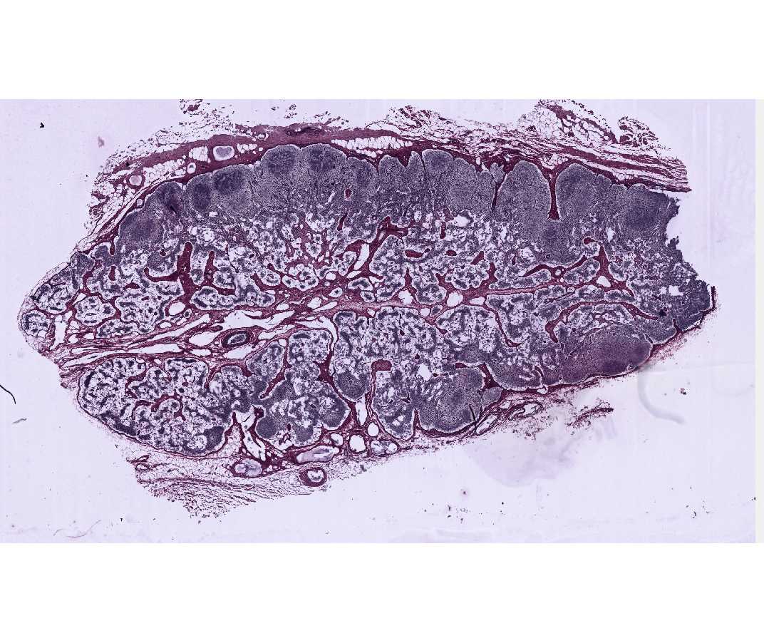
Identify the outer, collagenous connective tissue capsule surrounding the lymph node, and the trabeculae that penetrate into the interior of the node. These fibers are colored reddish-brown by the counterstain Azo-carmine. The black, silver-stained reticular fibers form the supporting framework for the cortical nodules. The lymphocytes, which are located within the interstices of this framework, are not well seen in this slide. The organization of lymph nodes will be studied in a future laboratory.