#8 – Healed Myocardial Infarct, with Myocardial Hypertrophy
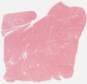
This slide comes from the post-mortem of the 56-year-old man described in slide #7. He had had multiple previous myocardial infarctions and was known to have multi-vessel coronary atherosclerosis, S/P bypass. At autopsy, there were multiple pale white-grey regions consistent with scars.
Broad, perivascular regions of fibrosis have replaced myocardium. The longitudinally-cut residual, viable myocytes show hypertrophy, characterised by fiber thickening and change of the nuclei from spindle – or cigar shaped – to “box car” like (rectangular). The fibrous tissue shows relatively few spindle cell nuclei of fibroblasts and large masses of pink collagen fibers.
Open with ImageScope
- boxcar nuclei in hypertrophied myocytes
- collagen fibers
- nuclei of fibroblasts
Regions of scarring after myocardial infarct impair conduction and may be associated with decreased cardiac contractility (with hypokinesis, ventricular dilatation, decreased ejection fraction if substantial) as well as increased incidence of arrhythmias. Demonstrate to your instructor / select a field for annotation showing: a region of fibrous scar (i.e., the site of previous cardiac myocytes which underwent necrosis).