SBPMD Histology Laboratory Manual
Coronary Atherosclerosis (slide #7)
Open with WebViewer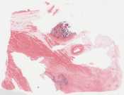
This 56-year-old man had a history of hypertension, hypercholesterolemia, type II diabetes mellitus and severe coronary artery disease, with prior surgery for multiple bypass grafts. He died of an acute myocardial infarction, characterised by acute chest pain with elevated cardiac enzymes (CK=485, LDH=216 and troponin 1=79.9)
This section shows epicardial fat, cross sections of the left anterior descending coronary artery (and several smaller arterial branches). The atherosclerotic plaque within the coronary artery shows several constituents, including cholesterol clefts, fibrosis and calcification. Note also the thrombotic material ( with lines of Zahn—platelets and fibrin alternating with red blood cells) which was found in the aneurysmal dilatation of the aortic sinus ( sinus of Valsalva). The wall around the thrombus shows haemorrhages and calcification.
Checklist: Have you identified
![]() Coronary Artery with plaque
Coronary Artery with plaque
![]() Cholesterol clefts
Cholesterol clefts
![]() Calcification
Calcification
![]() Fibrosis
Fibrosis
Questions
- What is the definition of “critical stenosis” of a coronary artery (the point at which symptomatic ischemic heart disease develops)?
- In addition to fixed atherosclerotic plaques in the coronary arteries, what are the other superimposed lesions that may play a role in developing myocardial ischemia?